
Chest CT Scan Analysis
A Chest Computed Tomography (CT) scan is an advanced imaging technique that provides highly detailed, cross-sectional views of your lungs and chest cavity. It is far more precise than a standard chest X-ray.
Do you need this procedure?
Understanding Chest CT Scans
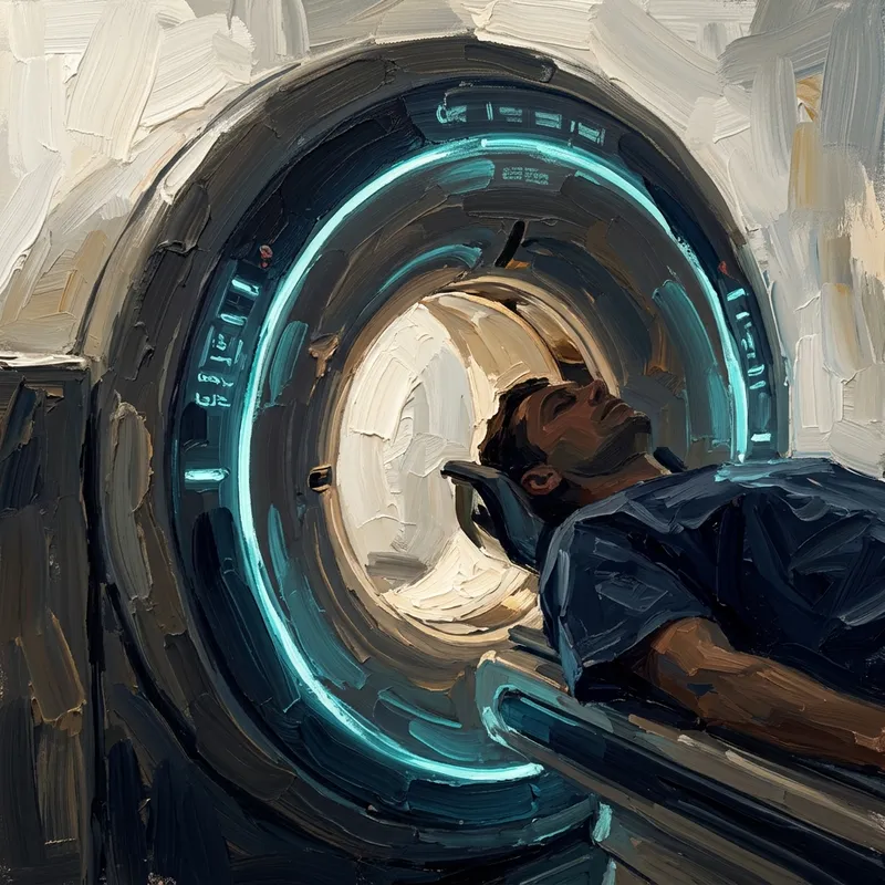
A CT scanner uses X-rays to take multiple pictures as it rotates around you. A computer then combines these images into detailed slices of your chest, allowing the doctor to view the internal structures of your lungs without overlapping tissues.
High-Resolution CT (HRCT) uses specific techniques to maximize the detail of the lung tissue, which is crucial for diagnosing subtle abnormalities.
When is a CT Scan Needed?
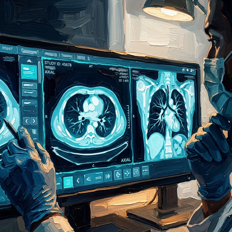
Chest CT scans are invaluable for detecting and managing serious lung conditions, including:
- Diagnosing Interstitial Lung Diseases (ILD) and pulmonary fibrosis.
- Detecting and staging lung cancer or other tumors.
- Evaluating complex respiratory infections or pneumonia that is not resolving.
- Investigating the cause of unexplained chronic cough or shortness of breath.
- Identifying pulmonary embolisms (blood clots in the lungs).
What to Expect
The scan is painless and usually takes less than 15 minutes. You will lie flat on a motorized table that slides into the donut-shaped scanner.
You may be asked to hold your breath for short periods to prevent blurring of the images. In some cases, a contrast dye may be injected into a vein in your arm to highlight specific blood vessels or structures.

Consult Dr. Feby Sadek for Chest CT Scan Analysis
Dr. Feby Sadek has extensive experience in diagnosing and treating chest conditions using the latest medical technologies. Book your consultation today to evaluate your condition and formulate the most suitable treatment plan.
Book your consultation now